|
|
|
Differential effects of recombinant fusion proteins TAT-OCT4 and TAT-NANOG on adult human fibroblasts |
Jiani CAO1,2, Zhifeng XIAO1, Bing CHEN1, Yuan GAO1, Chunying SHI1, Jinhuan WANG3, Jianwu DAI1( ) ) |
| 1. Key Laboratory of Molecular Developmental Biology, Institute of Genetics and Developmental Biology, Chinese Academy of Sciences, Beijing 100080, China; 2. Graduate School, Chinese Academy of Sciences, Beijing 100080, China; 3. Institute of Neurosurgery, Tianjin Huanhu Hospital, Tianjin 300060, China |
|
|
|
|
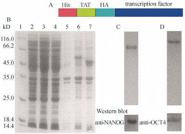
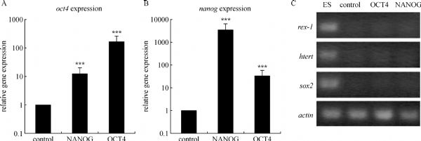
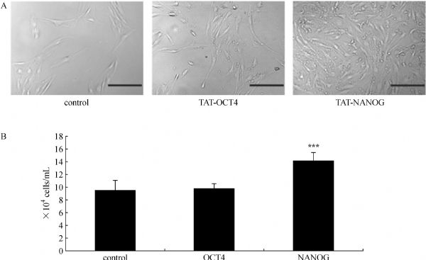
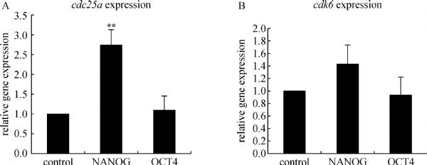
Abstract OCT4 and NANOG are two important transcription factors for maintaining the pluripotency and self-renewal abilities of embryonic stem (ES) cells. Meanwhile they play key roles in the induced pluripotent stem (iPS) cells. In this study, recombinant transcript factors TAT-NANOG and TAT-OCT4, which contained a fused powerful protein transduction domain (PTD) TAT from human immunodeficiency virus (HIV), were produced. Each fusion protein could be transported into human adult fibroblasts (HAF) successfully and activated the endogenous transcription of both nanog and oct4. Our study revealed the inter-regulation and autoregulation abilities of solo oct4 or nanog in the process of iPS cell reprogramming. Meanwhile the transduction of TAT-NANOG could accelerate the growth rate of HAF cells, and the key cell cycle regulator cdc25a was up-regulated. Thus cdc25a may be involved in the regulation of cell growth by NANOG. In addition, the TAT fusion protein technology provided a novel way to improve cell growth that is more controllable and safer.
|
| Keywords
induced pluripotent stem cells (iPS cells)
OCT4
NANOG
protein transduction domain
TAT
cell proliferation
|
|
Corresponding Author(s):
DAI Jianwu,Email:jwdai@genetics.ac.cn
|
|
Issue Date: 01 October 2010
|
|
| 1 |
Boyer L A, Lee T I, Cole M F, Johnstone S E, Levine S S, Zucker J P, Guenther M G, Kumar R M, Murray H L, Jenner R G, Gifford D K, Melton D A, Jaenisch R, Young R A (2005). Core transcriptional regulatory circuitry in human embryonic stem cells. Cell , 122(6): 947–956
|
| 2 |
Bradford M M (1976). A rapid and sensitive method for the quantitation of microgram quantities of protein utilizing the principle of protein-dye binding. Anal Biochem , 72: 248–254
doi: 10.1016/0003-2697(76)90527-3
|
| 3 |
Chambers I, Colby D, Robertson M, Nichols J, Lee S, Tweedie S, Smith A (2003). Functional expression cloning of nanog, a pluripotency sustaining factor in embryonic stem cells. Cell , 113(5): 643–655
|
| 4 |
Go M J, Takenaka C, Ohgushi H (2008). Forced expression of Sox2 or nanog in human bone marrow derived mesenchymal stem cells maintains their expansion and differentiation capabilities. Exp Cell Res , 314(5): 1147–1154
doi: 10.1016/j.yexcr.2007.11.021
|
| 5 |
Green M, Loewenstein P M (1988). Autonomous functional domains of chemically synthesized human immunodeficiency virus tat trans-activator protein. Cell , 55(6): 1179–1188
|
| 6 |
Hay D C, Sutherland L, Clark J, Burdon T (2004). Oct-4 knockdown induces similar patterns of endoderm and trophoblast differentiation markers in human and mouse embryonic stem cells. Stem Cells , 22(2): 225–235
doi: 10.1634/stemcells.22-2-225
|
| 7 |
Hough S R, Clements I, Welch P J, Wiederholt K A (2006). Differentiation of mouse embryonic stem cells after RNA interference-mediated silencing of OCT4 and nanog. Stem Cells , 24(6): 1467–1475
doi: 10.1634/stemcells.2005-0475
|
| 8 |
Hyslop L, Stojkovic M, Armstrong L, Walter T, Stojkovic P, Przyborski S, Herbert M, Murdoch A, Strachan T, Lako M (2005). Downregulation of NANOG induces differentiation of human embryonic stem cells to extraembryonic lineages. Stem Cells , 23(8): 1035–1043
doi: 10.1634/stemcells.2005-0080
|
| 9 |
Kim D, Kim C H, Moon J I, Chung Y G, Chang M Y, Han B S, Ko S, Yang E, Cha K Y, Lanza R, Kim K S (2009). Generation of human induced pluripotent stem cells by direct delivery of reprogramming proteins. Cell Stem Cell , 4(6): 472–476
doi: 10.1016/j.stem.2009.05.005
|
| 10 |
Loh Y H, Wu Q, Chew J L, Vega V B, Zhang W, Chen X, Bourque G, George J, Leong B, Liu J, Wong K Y, Sung K W, Lee C W, Zhao X D, Chiu K P, Lipovich L, Kuznetsov V A, Robson P, Stanton L W, Wei C L, Ruan Y, Lim B, Ng H H (2006). The oct4 and nanog transcription network regulates pluripotency in mouse embryonic stem cells. Nat Genet , 38(4): 431–440
doi: 10.1038/ng1760
|
| 11 |
Matin M M, Walsh J R, Gokhale P J, Draper J S, Bahrami A R, Morton I, Moore H D, Andrews P W (2004). Specific knockdown of oct4 and beta2-microglobulin expression by RNA interference in human embryonic stem cells and embryonic carcinoma cells. Stem Cells , 22(5): 659–668
doi: 10.1634/stemcells.22-5-659
|
| 12 |
Mitsui K, Tokuzawa Y, Itoh H, Segawa K, Murakami M, Takahashi K, Maruyama M, Maeda M, Yamanaka S (2003). The homeoprotein nanog is required for maintenance of pluripotency in mouse epiblast and ES cells. Cell , 113(5): 631–642
|
| 13 |
Nagahara H, Vocero-Akbani A M, Snyder E L, Ho A, Latham D G, Lissy N A, Becker-Hapak M, Ezhevsky S A, Dowdy S F (1998). Transduction of full-length TAT fusion proteins into mammalian cells: TAT-p27Kip1 induces cell migration. Nat Med , 4(12): 1449–1452
doi: 10.1038/4042
|
| 14 |
Nichols J, Zevnik B, Anastassiadis K, Niwa H, Klewe-Nebenius D, Chambers I, Sch?ler H, Smith A (1998). Formation of pluripotent stem cells in the mammalian embryo depends on the POU transcription factor oct4. Cell , 95(3): 379–391
|
| 15 |
Niwa H (2001). Molecular mechanism to maintain stem cell renewal of ES cells. Cell Struct Funct , 26(3): 137–148
doi: 10.1247/csf.26.137
|
| 16 |
Niwa H, Miyazaki J, Smith A G (2000). Quantitative expression of Oct-3/4 defines differentiation, dedifferentiation or self-renewal of ES cells. Nat Genet , 24(4): 372–376
doi: 10.1038/74199
|
| 17 |
Pesce M, Sch?ler H R (2001). Oct-4: gatekeeper in the beginnings of mammalian development. Stem Cells , 19(4): 271–278
doi: 10.1634/stemcells.19-4-271
|
| 18 |
Piestun D, Kochupurakkal B S, Jacob-Hirsch J, Zeligson S, Koudritsky M, Domany E, Amariglio N, Rechavi G, Givol D (2006). nanog transforms NIH3T3 cells and targets cell-type restricted genes. Biochem Biophys Res Commun , 343(1): 279–285
doi: 10.1016/j.bbrc.2006.02.152
|
| 19 |
Schwarze S R, Ho A, Vocero-Akbani A, Dowdy S F (1999). In vivo protein transduction: delivery of a biologically active protein into the mouse. Science , 285(5433): 1569–1572
doi: 10.1126/science.285.5433.1569
|
| 20 |
Silva J, Nichols J, Theunissen T W, Guo G, van Oosten A L, Barrandon O, Wray J, Yamanaka S, Chambers I, Smith A (2009). nanog is the gateway to the pluripotent ground state. Cell , 138(4): 722–737
|
| 21 |
Takahashi K, Tanabe K, Ohnuki M, Narita M, Ichisaka T, Tomoda K, Yamanaka S (2007). Induction of pluripotent stem cells from adult human fibroblasts by defined factors. Cell , 131(5): 861–872
|
| 22 |
Takahashi K, Yamanaka S (2006). Induction of pluripotent stem cells from mouse embryonic and adult fibroblast cultures by defined factors. Cell , 126(4): 663–676
|
| 23 |
Wang J, Rao S, Chu J, Shen X, Levasseur D N, Theunissen T W, Orkin S H (2006). A protein interaction network for pluripotency of embryonic stem cells. Nature , 444(7117): 364–368
doi: 10.1038/nature05284
|
| 24 |
Yu J, Hu K, Smuga-Otto K, Tian S, Stewart R, Slukvin I I, Thomson J A (2009). Human induced pluripotent stem cells free of vector and transgene sequences. Science , 324(5928): 797–801
doi: 10.1126/science.1172482
|
| 25 |
Yu J, Vodyanik M A, Smuga-Otto K, Antosiewicz-Bourget J, Frane J L, Tian S, Nie J, Jonsdottir G A, Ruotti V, Stewart R, Slukvin II, Thomson J A (2007). Induced pluripotent stem cell lines derived from human somatic cells. Science , 318, 1917–1920
doi: 10.1126/science.1151526
|
| 26 |
Zaehres H, Lensch M W, Daheron L, Stewart S A, Itskovitz-Eldor J, Daley G Q (2005). High-efficiency RNA interference in human embryonic stem cells. Stem Cells , 23(3): 299–305
doi: 10.1634/stemcells.2004-0252
|
| 27 |
Zhang J, Wang X, Chen B, Suo G, Zhao Y, Duan Z, Dai J (2005). Expression of nanog gene promotes NIH3T3 cell proliferation. Biochem Biophys Res Commun , 338(2): 1098–1102
doi: 10.1016/j.bbrc.2005.10.071
|
| 28 |
Zhang X, Neganova I, Przyborski S, Yang C, Cooke M, Atkinson S P, Anyfantis G, Fenyk S, Keith W N, Hoare S F, Hughes O, Strachan T, Stojkovic M, Hinds P W, Armstrong L, Lako M (2009). A role for NANOG in G1 to S transition in human embryonic stem cells through direct binding of CDK6 and cdc25a. J Cell Biol , 184(1): 67–82
doi: 10.1083/jcb.200801009
|
| 29 |
Zhou H, Wu S, Joo J Y, Zhu S, Han D W, Lin T, Trauger S, Bien G, Yao S, Zhu Y, Siuzdak G, Sch?ler H R, Duan L, Ding S (2009). Generation of induced pluripotent stem cells using recombinant proteins. Cell Stem Cell , 4(5): 381–384
doi: 10.1016/j.stem.2009.04.005
|
|
Viewed |
|
|
|
Full text
|
|
|
|
|
Abstract
|
|
|
|
|
Cited |
|
|
|
|
| |
Shared |
|
|
|
|
| |
Discussed |
|
|
|
|