|
|
|
Immunosuppressive tumor microenvironment contributes to tumor progression in diffuse large B-cell lymphoma upon anti-CD19 chimeric antigen receptor T therapy |
Zixun Yan1, Li Li1,5, Di Fu1, Wen Wu1, Niu Qiao1, Yaohui Huang1, Lu Jiang1, Depei Wu2, Yu Hu3, Huilai Zhang4, Pengpeng Xu1, Shu Cheng1, Li Wang1, Sahin Lacin5, Muharrem Muftuoglu5, Weili Zhao1,6( ) ) |
1. Shanghai Institute of Hematology, State Key Laboratory of Medical Genomics, National Research Center for Translational Medicine at Shanghai, Ruijin Hospital Affiliated to Shanghai Jiao Tong University School of Medicine, Shanghai 200025, China
2. Institute of Hematology, The First Affiliated Hospital of Soochow University, Suzhou 215000, China
3. Institute of Hematology, Union Hospital, Tongji Medical College, Huazhong University of Science and Technology, Wuhan 430000, China
4. Tianjin Medical University Cancer Institute & Hospital, Tianjin 300070, China
5. University of Texas MD Anderson Cancer Center, Houston, Texas 77030, USA
6. Pôle de Recherches Sino-Français en Science du Vivant et Génomique, Laboratory of Molecular Pathology, Shanghai 200025, China |
|
|
|
|
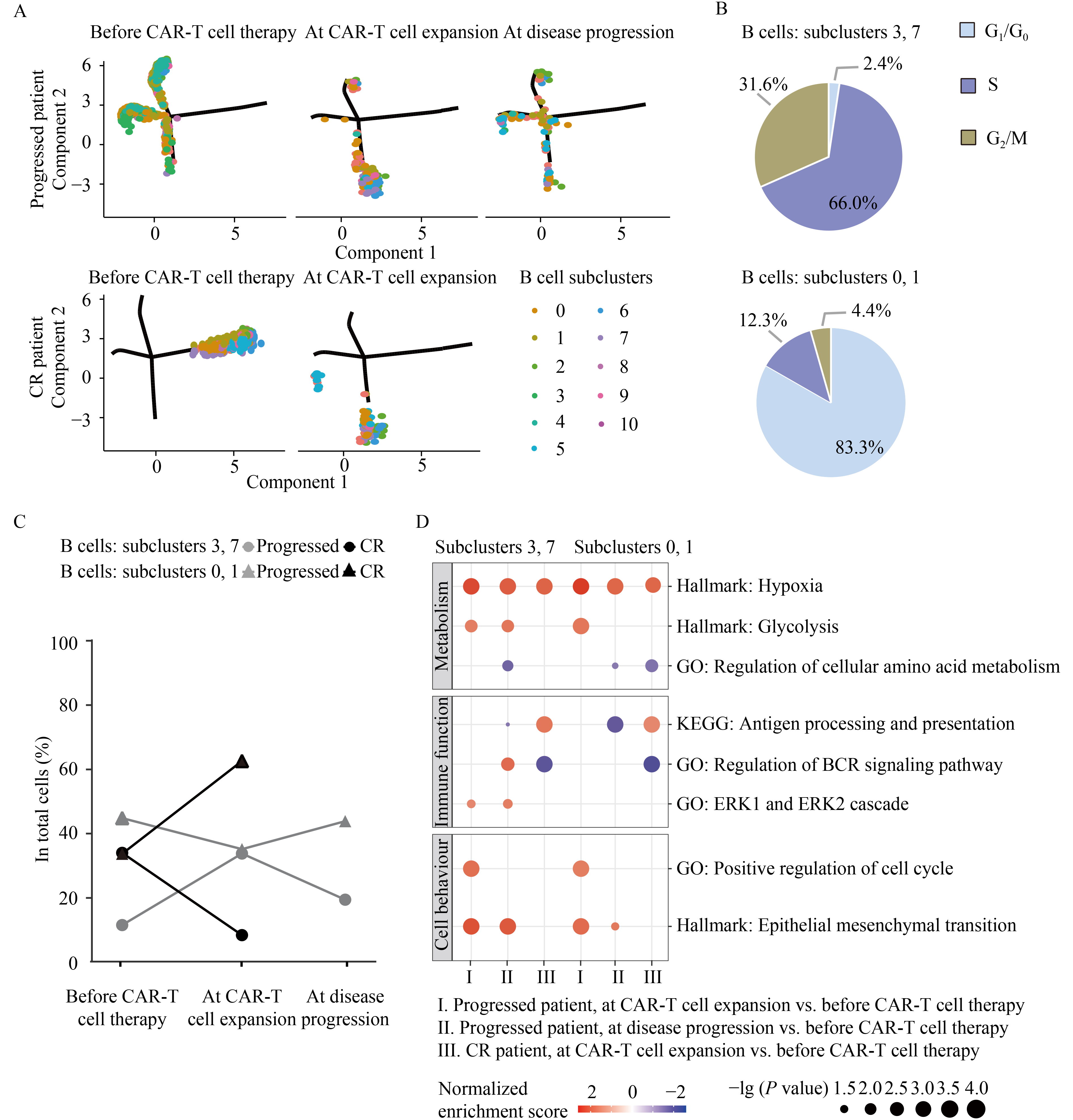
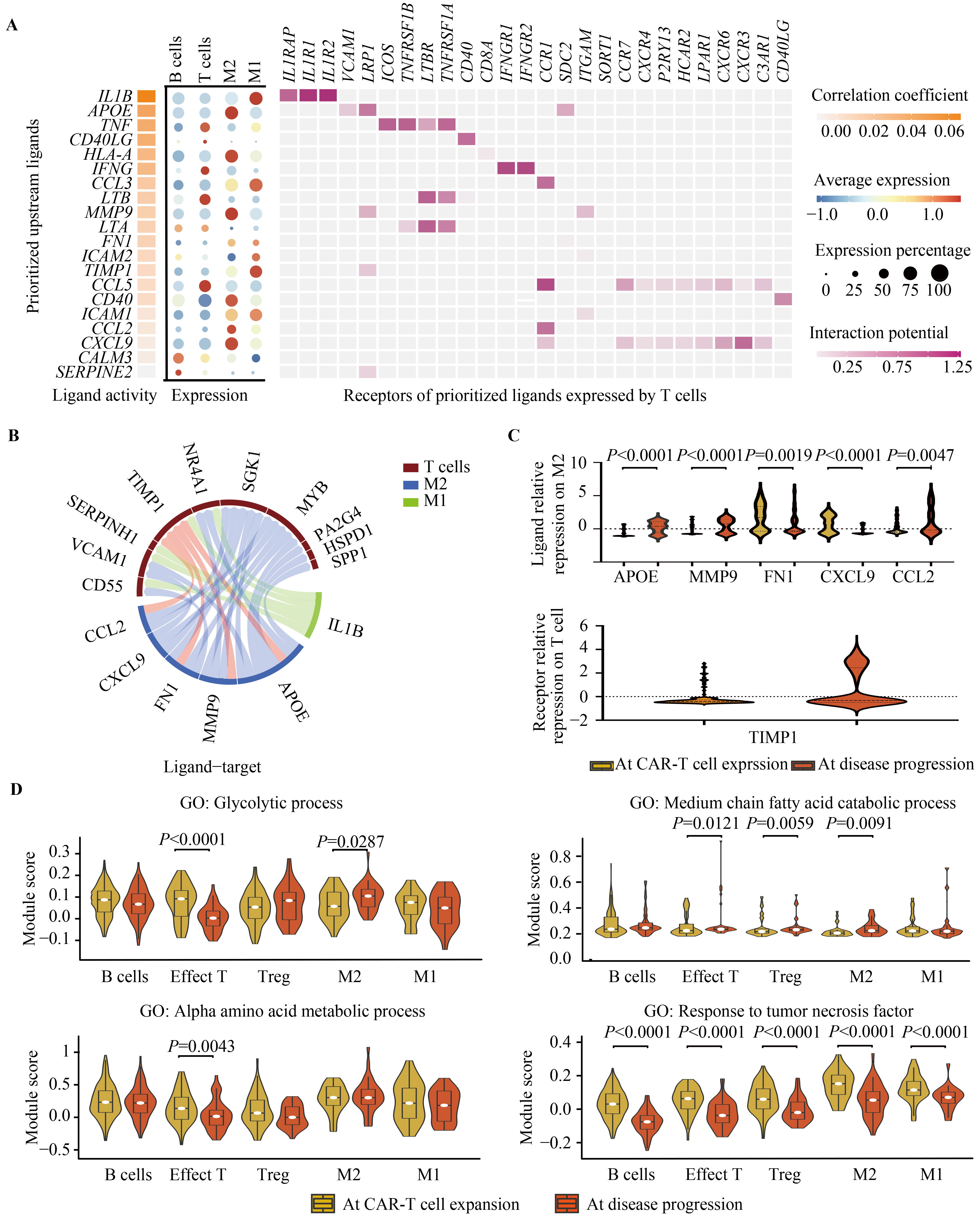
Abstract Anti-CD19 chimeric antigen receptor (CAR)-T cell therapy has achieved 40%–50% long-term complete response in relapsed or refractory diffuse large B-cell lymphoma (DLBCL) patients. However, the underlying mechanism of alterations in the tumor microenvironments resulting in CAR-T cell therapy failure needs further investigation. A multi-center phase I/II trial of anti-CD19 CD28z CAR-T (FKC876, ChiCTR1800019661) was conducted. Among 22 evaluable DLBCL patients, seven achieved complete remission, 10 experienced partial remissions, while four had stable disease by day 29. Single-cell RNA sequencing results were obtained from core needle biopsy tumor samples collected from long-term complete remission and early-progressed patients, and compared at different stages of treatment. M2-subtype macrophages were significantly involved in both in vivo and in vitro anti-tumor functions of CAR-T cells, leading to CAR-T cell therapy failure and disease progression in DLBCL. Immunosuppressive tumor microenvironments persisted before CAR-T cell therapy, during both cell expansion and disease progression, which could not be altered by infiltrating CAR-T cells. Aberrant metabolism profile of M2-subtype macrophages and those of dysfunctional T cells also contributed to the immunosuppressive tumor microenvironments. Thus, our findings provided a clinical rationale for targeting tumor microenvironments and reprogramming immune cell metabolism as effective therapeutic strategies to prevent lymphoma relapse in future designs of CAR-T cell therapy.
|
| Keywords
anti-CD19 chimeric antigen receptor T
immunotherapy
diffuse large B cell lymphoma
tumor microenvironment
tumor-associated macrophage
metabolism
|
|
Corresponding Author(s):
Weili Zhao
|
|
Just Accepted Date: 07 December 2022
Online First Date: 17 April 2023
Issue Date: 12 October 2023
|
|
| 1 |
JH Park, I Rivière, M Gonen, X Wang, B Sénéchal, KJ Curran, C Sauter, Y Wang, B Santomasso, E Mead, M Roshal, P Maslak, M Davila, RJ Brentjens, M Sadelain. Long-term follow-up of CD19 CAR therapy in acute lymphoblastic leukemia. N Engl J Med 2018; 378(5): 449–459
https://doi.org/10.1056/NEJMoa1709919
pmid: 29385376
|
| 2 |
SJ Schuster, MR Bishop, CS Tam, EK Waller, P Borchmann, JP McGuirk, U Jäger, S Jaglowski, C Andreadis, JR Westin, I Fleury, V Bachanova, SR Foley, PJ Ho, S Mielke, JM Magenau, H Holte, S Pantano, LB Pacaud, R Awasthi, J Chu, Ö Anak, G Salles, RT; JULIET Investigators Maziarz. Tisagenlecleucel in adult relapsed or refractory diffuse large B-cell lymphoma. N Engl J Med 2019; 380(1): 45–56
https://doi.org/10.1056/NEJMoa1804980
pmid: 30501490
|
| 3 |
SS Neelapu, FL Locke, NL Bartlett, LJ Lekakis, DB Miklos, CA Jacobson, I Braunschweig, OO Oluwole, T Siddiqi, Y Lin, JM Timmerman, PJ Stiff, JW Friedberg, IW Flinn, A Goy, BT Hill, MR Smith, A Deol, U Farooq, P McSweeney, J Munoz, I Avivi, JE Castro, JR Westin, JC Chavez, A Ghobadi, KV Komanduri, R Levy, ED Jacobsen, TE Witzig, P Reagan, A Bot, J Rossi, L Navale, Y Jiang, J Aycock, M Elias, D Chang, J Wiezorek, WY Go. Axicabtagene ciloleucel CAR T-cell therapy in refractory large B-Cell lymphoma. N Engl J Med 2017; 377(26): 2531–2544
https://doi.org/10.1056/NEJMoa1707447
pmid: 29226797
|
| 4 |
N Singh, YG Lee, O Shestova, P Ravikumar, KE Hayer, SJ Hong, XM Lu, R Pajarillo, S Agarwal, S Kuramitsu, EJ Orlando, KT Mueller, CR Good, SL Berger, O Shalem, MD Weitzman, NV Frey, SL Maude, SA Grupp, CH June, S Gill, M Ruella. Impaired death receptor signaling in leukemia causes antigen-independent resistance by inducing CAR T-cell dysfunction. Cancer Discov 2020; 10(4): 552–567
https://doi.org/10.1158/2159-8290.CD-19-0813
pmid: 32001516
|
| 5 |
EJ Orlando, X Han, C Tribouley, PA Wood, RJ Leary, M Riester, JE Levine, M Qayed, SA Grupp, M Boyer, B De Moerloose, ER Nemecek, H Bittencourt, H Hiramatsu, J Buechner, SM Davies, MR Verneris, K Nguyen, JL Brogdon, H Bitter, M Morrissey, P Pierog, S Pantano, JA Engelman, W Winckler. Genetic mechanisms of target antigen loss in CAR19 therapy of acute lymphoblastic leukemia. Nat Med 2018; 24(10): 1504–1506
https://doi.org/10.1038/s41591-018-0146-z
pmid: 30275569
|
| 6 |
E Sotillo, DM Barrett, KL Black, A Bagashev, D Oldridge, G Wu, R Sussman, C Lanauze, M Ruella, MR Gazzara, NM Martinez, CT Harrington, EY Chung, J Perazzelli, TJ Hofmann, SL Maude, P Raman, A Barrera, S Gill, SF Lacey, JJ Melenhorst, D Allman, E Jacoby, T Fry, C Mackall, Y Barash, KW Lynch, JM Maris, SA Grupp, A Thomas-Tikhonenko. Convergence of acquired mutations and alternative splicing of CD19 enables resistance to CART-19 immunotherapy. Cancer Discov 2015; 5(12): 1282–1295
https://doi.org/10.1158/2159-8290.CD-15-1020
pmid: 26516065
|
| 7 |
CJ Turtle, LA Hanafi, C Berger, TA Gooley, S Cherian, M Hudecek, D Sommermeyer, K Melville, B Pender, TM Budiarto, E Robinson, NN Steevens, C Chaney, L Soma, X Chen, C Yeung, B Wood, D Li, J Cao, S Heimfeld, MC Jensen, SR Riddell, DG Maloney. CD19 CAR-T cells of defined CD4+:CD8+ composition in adult B cell ALL patients. J Clin Invest 2016; 126(6): 2123–2138
https://doi.org/10.1172/JCI85309
pmid: 27111235
|
| 8 |
JA Fraietta, SF Lacey, EJ Orlando, I Pruteanu-Malinici, M Gohil, S Lundh, AC Boesteanu, Y Wang, RS O’Connor, WT Hwang, E Pequignot, DE Ambrose, C Zhang, N Wilcox, F Bedoya, C Dorfmeier, F Chen, L Tian, H Parakandi, M Gupta, RM Young, FB Johnson, I Kulikovskaya, L Liu, J Xu, SH Kassim, MM Davis, BL Levine, NV Frey, DL Siegel, AC Huang, EJ Wherry, H Bitter, JL Brogdon, DL Porter, CH June, JJ Melenhorst. Determinants of response and resistance to CD19 chimeric antigen receptor (CAR) T cell therapy of chronic lymphocytic leukemia. Nat Med 2018; 24(5): 563–571
https://doi.org/10.1038/s41591-018-0010-1
pmid: 29713085
|
| 9 |
ZX Yan, L Li, W Wang, BS OuYang, S Cheng, L Wang, W Wu, PP Xu, M Muftuoglu, M Hao, S Yang, MC Zhang, Z Zheng, J Li, WL Zhao. Clinical efficacy and tumor microenvironment influence in a dose-escalation study of anti-CD19 chimeric antigen receptor T cells in refractory B-cell non-Hodgkin’s lymphoma. Clin Cancer Res 2019; 25(23): 6995–7003
https://doi.org/10.1158/1078-0432.CCR-19-0101
pmid: 31444250
|
| 10 |
H Zola, PJ MacArdle, T Bradford, H Weedon, H Yasu, Y Kurosawa. Preparation and characterization of a chimeric CD19 monoclonal antibody. Immunol Cell Biol 1991; 69(6): 411–422
https://doi.org/10.1038/icb.1991.58
pmid: 1725979
|
| 11 |
R Shen, PP Xu, N Wang, HM Yi, L Dong, D Fu, JY Huang, HY Huang, A Janin, S Cheng, L Wang, WL Zhao. Influence of oncogenic mutations and tumor microenvironment alterations on extranodal invasion in diffuse large B-cell lymphoma. Clin Transl Med 2020; 10(7): e221
https://doi.org/10.1002/ctm2.221
pmid: 33252851
|
| 12 |
SL Maude, TW Laetsch, J Buechner, S Rives, M Boyer, H Bittencourt, P Bader, MR Verneris, HE Stefanski, GD Myers, M Qayed, B De Moerloose, H Hiramatsu, K Schlis, KL Davis, PL Martin, ER Nemecek, GA Yanik, C Peters, A Baruchel, N Boissel, F Mechinaud, A Balduzzi, J Krueger, CH June, BL Levine, P Wood, T Taran, M Leung, KT Mueller, Y Zhang, K Sen, D Lebwohl, MA Pulsipher, SA Grupp. Tisagenlecleucel in children and young adults with B-cell lymphoblastic leukemia. N Engl J Med 2018; 378(5): 439–448
https://doi.org/10.1056/NEJMoa1709866
pmid: 29385370
|
| 13 |
C Feig, JO Jones, M Kraman, RJ Wells, A Deonarine, DS Chan, CM Connell, EW Roberts, Q Zhao, OL Caballero, SA Teichmann, T Janowitz, DI Jodrell, DA Tuveson, DT Fearon. Targeting CXCL12 from FAP-expressing carcinoma-associated fibroblasts synergizes with anti-PD-L1 immunotherapy in pancreatic cancer. Proc Natl Acad Sci USA 2013; 110(50): 20212–20217
https://doi.org/10.1073/pnas.1320318110
pmid: 24277834
|
| 14 |
J Rossi, P Paczkowski, YW Shen, K Morse, B Flynn, A Kaiser, C Ng, K Gallatin, T Cain, R Fan, S Mackay, JR Heath, SA Rosenberg, JN Kochenderfer, J Zhou, A Bot. Preinfusion polyfunctional anti-CD19 chimeric antigen receptor T cells are associated with clinical outcomes in NHL. Blood 2018; 132(8): 804–814
https://doi.org/10.1182/blood-2018-01-828343
pmid: 29895668
|
| 15 |
D Lee, D Hokinson, S Park, R Elvira, F Kusuma, JM Lee, M Yun, SG Lee, J Han. ER stress induces cell cycle arrest at the G2/M phase through eIF2α phosphorylation and GADD45α. Int J Mol Sci 2019; 20(24): 6309
https://doi.org/10.3390/ijms20246309
pmid: 31847234
|
| 16 |
G Song, S Xu, H Zhang, Y Wang, C Xiao, T Jiang, L Wu, T Zhang, X Sun, L Zhong, C Zhou, Z Wang, Z Peng, J Chen, X Wang. TIMP1 is a prognostic marker for the progression and metastasis of colon cancer through FAK-PI3K/AKT and MAPK pathway. J Exp Clin Cancer Res 2016; 35(1): 148
https://doi.org/10.1186/s13046-016-0427-7
pmid: 27644693
|
| 17 |
RD Leone, JD Powell. Metabolism of immune cells in cancer. Nat Rev Cancer 2020; 20(9): 516–531
https://doi.org/10.1038/s41568-020-0273-y
pmid: 32632251
|
| 18 |
AAJ Hamers, AB Pillai. A sweet alternative: maintaining M2 macrophage polarization. Sci Immunol 2018; 3(29): eaav7759
https://doi.org/10.1126/sciimmunol.aav7759
pmid: 30389801
|
| 19 |
B Faubert, A Solmonson, RJ DeBerardinis. Metabolic reprogramming and cancer progression. Science 2020; 368(6487): eaaw5473
https://doi.org/10.1126/science.aaw5473
pmid: 32273439
|
| 20 |
I Vitale, G Manic, LM Coussens, G Kroemer, L Galluzzi. Macrophages and metabolism in the tumor microenvironment. Cell Metab 2019; 30(1): 36–50
https://doi.org/10.1016/j.cmet.2019.06.001
pmid: 31269428
|
| 21 |
R Eil, SK Vodnala, D Clever, CA Klebanoff, M Sukumar, JH Pan, DC Palmer, A Gros, TN Yamamoto, SJ Patel, GC Guittard, Z Yu, V Carbonaro, K Okkenhaug, DS Schrump, WM Linehan, R Roychoudhuri, NP Restifo. Ionic immune suppression within the tumour microenvironment limits T cell effector function. Nature 2016; 537(7621): 539–543
https://doi.org/10.1038/nature19364
pmid: 27626381
|
| 22 |
Z Xiao, Z Dai, JW Locasale. Metabolic landscape of the tumor microenvironment at single cell resolution. Nat Commun 2019; 10(1): 3763
https://doi.org/10.1038/s41467-019-11738-0
pmid: 31434891
|
| 23 |
MS Nakazawa, B Keith, MC Simon. Oxygen availability and metabolic adaptations. Nat Rev Cancer 2016; 16(10): 663–673
https://doi.org/10.1038/nrc.2016.84
pmid: 27658636
|
| 24 |
CH Chang, JD Curtis, LB Jr Maggi, B Faubert, AV Villarino, D O’Sullivan, SC Huang, der Windt GJ van, J Blagih, J Qiu, JD Weber, EJ Pearce, RG Jones, EL Pearce. Posttranscriptional control of T cell effector function by aerobic glycolysis. Cell 2013; 153(6): 1239–1251
https://doi.org/10.1016/j.cell.2013.05.016
pmid: 23746840
|
| 25 |
CM Cham, TF Gajewski. Glucose availability regulates IFN-γ production and p70S6 kinase activation in CD8+ effector T cells. J Immunol 2005; 174(8): 4670–4677
https://doi.org/10.4049/jimmunol.174.8.4670
pmid: 15814691
|
| 26 |
L Berod, C Friedrich, A Nandan, J Freitag, S Hagemann, K Harmrolfs, A Sandouk, C Hesse, CN Castro, H Bähre, SK Tschirner, N Gorinski, M Gohmert, CT Mayer, J Huehn, E Ponimaskin, WR Abraham, R Müller, M Lochner, T Sparwasser. De novo fatty acid synthesis controls the fate between regulatory T and T helper 17 cells. Nat Med 2014; 20(11): 1327–1333
https://doi.org/10.1038/nm.3704
pmid: 25282359
|
| 27 |
Q Deng, G Han, N Puebla-Osorio, MCJ Ma, P Strati, B Chasen, E Dai, M Dang, N Jain, H Yang, Y Wang, S Zhang, R Wang, R Chen, J Showell, S Ghosh, S Patchva, Q Zhang, R Sun, F Hagemeister, L Fayad, F Samaniego, HC Lee, LJ Nastoupil, N Fowler, R Eric Davis, J Westin, SS Neelapu, L Wang, MR Green. Characteristics of anti-CD19 CAR T cell infusion products associated with efficacy and toxicity in patients with large B cell lymphomas. Nat Med 2020; 26(12): 1878–1887
https://doi.org/10.1038/s41591-020-1061-7
pmid: 33020644
|
| 28 |
CH Chang, J Qiu, D O’Sullivan, MD Buck, T Noguchi, JD Curtis, Q Chen, M Gindin, MM Gubin, der Windt GJ van, E Tonc, RD Schreiber, EJ Pearce, EL Pearce. Metabolic competition in the tumor microenvironment is a driver of cancer progression. Cell 2015; 162(6): 1229–1241
https://doi.org/10.1016/j.cell.2015.08.016
pmid: 26321679
|
|
Viewed |
|
|
|
Full text
|
|
|
|
|
Abstract
|
|
|
|
|
Cited |
|
|
|
|
| |
Shared |
|
|
|
|
| |
Discussed |
|
|
|
|