|
|
|
Technology developments and biomedical applications of polarization-sensitive optical coherence tomography |
Zhenyang DING,Chia-Pin LIANG,Yu CHEN( ) ) |
| Fischell Department of Bioengineering, University of Maryland, College Park, MD 20742, USA |
|
|
|
|
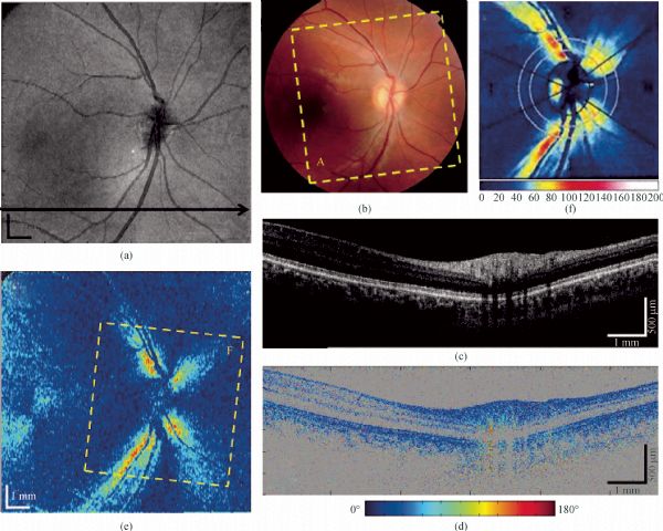
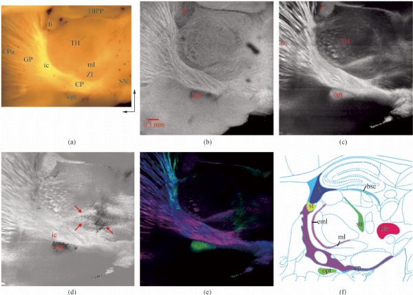
Abstract Polarization-sensitive optical coherence tomography (PS-OCT) enables depth-resolved mapping of sample polarization information, such as phase-retardation and optical axis orientation, which is particularly useful when the nano-scale organization of tissue that are difficult to be observed in the intensity images of a regular optical coherence tomography (OCT). In this review, we survey two types of methods and systems of PS-OCT. The first type is PS-OCT with single input polarization state, which contain bulk optics or polarization maintaining fiber (PMF) based systems and single-mode fiber (SMF) based systems. The second type is PS-OCT with two different input polarization states, which contain SMF based systems and PMF based systems, through either time, frequency, or depth multiplexing. In addition, representative biomedical applications using PS-OCT, such as retinal imaging, skin cancer detection, and brain mapping, are demonstrated.
|
| Keywords
optical coherence tomography (OCT)
polarization-sensitive optical coherence tomography (PS-OCT)
polarization
imaging
|
|
Corresponding Author(s):
Yu CHEN
|
|
Just Accepted Date: 04 February 2015
Issue Date: 24 June 2015
|
|
| 1 |
Hee M R, Huang D, Swanson E A, Fujimoto J G. Polarization-sensitive low-coherence reflectometer for birefringence characterization and ranging. Journal of the Optical Society of America. B, Optical Physics, 1992, 9(6): 903–908
https://doi.org/10.1364/JOSAB.9.000903
|
| 2 |
Huang D, Swanson E A, Lin C P, Schuman J S, Stinson W G, Chang W, Hee M R, Flotte T, Gregory K, Puliafito C A. Optical coherence tomography. Science, 1991, 254(5035): 1178–1181
https://doi.org/10.1126/science.1957169
pmid: 1957169
|
| 3 |
de Boer J, Srinivas S, Malekafzali A, Chen Z, Nelson J. Imaging thermally damaged tissue by polarization sensitive optical coherence tomography. Optics Express, 1998, 3(6): 212–218
https://doi.org/10.1364/OE.3.000212
pmid: 19384363
|
| 4 |
Schoenenberger K, Colston B W, Maitland D J, Da Silva L B, Everett M J. Mapping of birefringence and thermal damage in tissue by use of polarization-sensitive optical coherence tomography. Applied Optics, 1998, 37(25): 6026–6036
https://doi.org/10.1364/AO.37.006026
pmid: 18286100
|
| 5 |
Park B H, Saxer C, Srinivas S M, Nelson J S, de Boer J F. In vivo burn depth determination by high-speed fiber-based polarization sensitive optical coherence tomography. Journal of Biomedical Optics, 2001, 6(4): 474–479
https://doi.org/10.1117/1.1413208
pmid: 11728208
|
| 6 |
Jiao S, Wang L V. Jones-matrix imaging of biological tissues with quadruple-channel optical coherence tomography. Journal of Biomedical Optics, 2002, 7(3): 350–358
https://doi.org/10.1117/1.1483878
pmid: 12175284
|
| 7 |
Jiao S, Yu W, Stoica G, Wang L V. Contrast mechanisms in polarization-sensitive Mueller-matrix optical coherence tomography and application in burn imaging. Applied Optics, 2003, 42(25): 5191–5197
https://doi.org/10.1364/AO.42.005191
pmid: 12962400
|
| 8 |
Pierce M C, Strasswimmer J, Park B H, Cense B, de Boer J F. Advances in optical coherence tomography imaging for dermatology. The Journal of Investigative Dermatology, 2004, 123(3): 458–463
https://doi.org/10.1111/j.0022-202X.2004.23404.x
pmid: 15304083
|
| 9 |
Srinivas S M, de Boer J F, Park H, Keikhanzadeh K, Huang H E, Zhang J, Jung W Q, Chen Z, Nelson J S. Determination of burn depth by polarization-sensitive optical coherence tomography. Journal of Biomedical Optics, 2004, 9(1): 207–212
https://doi.org/10.1117/1.1629680
pmid: 14715075
|
| 10 |
Strasswimmer J, Pierce M C, Park B H, Neel V, de Boer J F. Polarization-sensitive optical coherence tomography of invasive basal cell carcinoma. Journal of Biomedical Optics, 2004, 9(2): 292–298
https://doi.org/10.1117/1.1644118
pmid: 15065894
|
| 11 |
Duan L, Marvdashti T, Lee A, Tang J Y, Ellerbee A K. Automated identification of basal cell carcinoma by polarization-sensitive optical coherence tomography. Biomedical Optics Express, 2014, 5(10): 3717–3729
https://doi.org/10.1364/BOE.5.003717
pmid: 25360384
|
| 12 |
Pircher M, Goetzinger E, Leitgeb R, Hitzenberger C K. Transversal phase resolved polarization sensitive optical coherence tomography. Physics in Medicine and Biology, 2004, 49(7): 1257–1263
https://doi.org/10.1088/0031-9155/49/7/013
pmid: 15128203
|
| 13 |
G?tzinger E, Pircher M, Sticker M, Fercher A F, Hitzenberger C K. Measurement and imaging of birefringent properties of the human cornea with phase-resolved, polarization-sensitive optical coherence tomography. Journal of Biomedical Optics, 2004, 9(1): 94–102
https://doi.org/10.1117/1.1629308
pmid: 14715060
|
| 14 |
Ducros M G, de Boer J F, Huang H E, Chao L C, Chen Z P, Nelson J S, Milner T E, Rylander H III. Polarization sensitive optical coherence tomography of the rabbit eye. IEEE Journal on Selected Topics in Quantum Electronics, 1999, 5(4): 1159–1167
https://doi.org/10.1109/2944.796342
|
| 15 |
Ducros M G, Marsack J D, Rylander H G 3rd, Thomsen S L, Milner T E. Primate retina imaging with polarization-sensitive optical coherence tomography. Journal of the Optical Society of America A, Optics, Image Science, and Vision, 2001, 18(12): 2945–2956
https://doi.org/10.1364/JOSAA.18.002945
pmid: 11760194
|
| 16 |
Cense B, Chen T C, Park B H, Pierce M C, de Boer J F. In vivo birefringence and thickness measurements of the human retinal nerve fiber layer using polarization-sensitive optical coherence tomography. Journal of Biomedical Optics, 2004, 9(1): 121–125
https://doi.org/10.1117/1.1627774
pmid: 14715063
|
| 17 |
G?tzinger E, Pircher M, Hitzenberger C K. High speed spectral domain polarization sensitive optical coherence tomography of the human retina. Optics Express, 2005, 13(25): 10217–10229
https://doi.org/10.1364/OPEX.13.010217
pmid: 19503236
|
| 18 |
Kemp N J, Park J, Zaatari H N, Rylander H G, Milner T E. High-sensitivity determination of birefringence in turbid media with enhanced polarization-sensitive optical coherence tomography. Journal of the Optical Society of America A, Optics, Image Science, and Vision, 2005, 22(3): 552–560
https://doi.org/10.1364/JOSAA.22.000552
pmid: 15770994
|
| 19 |
Baumann B, Choi W, Potsaid B, Huang D, Duker J S, Fujimoto J G. Swept source/Fourier domain polarization sensitive optical coherence tomography with a passive polarization delay unit. Optics Express, 2012, 20(9): 10229–10241
https://doi.org/10.1364/OE.20.010229
pmid: 22535114
|
| 20 |
G?tzinger E, Pircher M, Geitzenauer W, Ahlers C, Baumann B, Michels S, Schmidt-Erfurth U, Hitzenberger C K. Retinal pigment epithelium segmentation by polarization sensitive optical coherence tomography. Optics Express, 2008, 16(21): 16410–16422
https://doi.org/10.1364/OE.16.016410
pmid: 18852747
|
| 21 |
Zotter S, Pircher M, Torzicky T, Baumann B, Yoshida H, Hirose F, Roberts P, Ritter M, Schütze C, G?tzinger E, Trasischker W, Vass C, Schmidt-Erfurth U, Hitzenberger C K. Large-field high-speed polarization sensitive spectral domain OCT and its applications in ophthalmology. Biomedical Optics Express, 2012, 3(11): 2720–2732
https://doi.org/10.1364/BOE.3.002720
pmid: 23162711
|
| 22 |
Cense B, Chen T C, Park B H, Pierce M C, de Boer J F. Thickness and birefringence of healthy retinal nerve fiber layer tissue measured with polarization-sensitive optical coherence tomography. Investigative Ophthalmology & Visual Science, 2004, 45(8): 2606–2612
https://doi.org/10.1167/iovs.03-1160
pmid: 15277483
|
| 23 |
Makita S, Yamanari M, Yasuno Y. Generalized Jones matrix optical coherence tomography: performance and local birefringence imaging. Optics Express, 2010, 18(2): 854–876
https://doi.org/10.1364/OE.18.000854
pmid: 20173907
|
| 24 |
Wang X J, Milner T E, de Boer J F, Zhang Y, Pashley D H, Nelson J S. Characterization of dentin and enamel by use of optical coherence tomography. Applied Optics, 1999, 38(10): 2092–2096
https://doi.org/10.1364/AO.38.002092
pmid: 18319769
|
| 25 |
Baumgartner A, Dichtl S, Hitzenberger C K, Sattmann H, Robl B, Moritz A, Fercher A F, Sperr W. Polarization-sensitive optical coherence tomography of dental structures. Caries Research, 2000, 34(1): 59–69
https://doi.org/10.1159/000016571
pmid: 10601786
|
| 26 |
Fried D, Xie J, Shafi S, Featherstone J D, Breunig T M, Le C. Imaging caries lesions and lesion progression with polarization sensitive optical coherence tomography. Journal of Biomedical Optics, 2002, 7(4): 618–627
https://doi.org/10.1117/1.1509752
pmid: 12421130
|
| 27 |
Chen Y, Otis L, Piao D, Zhu Q. Characterization of dentin, enamel, and carious lesions by a polarization-sensitive optical coherence tomography system. Applied Optics, 2005, 44(11): 2041–2048
https://doi.org/10.1364/AO.44.002041
pmid: 15835353
|
| 28 |
Jones R S, Darling C L, Featherstone J D, Fried D. Remineralization of in vitro dental caries assessed with polarization-sensitive optical coherence tomography. Journal of biomedical optics, 2006, 11(1): 014016
|
| 29 |
Pierce M, Shishkov M, Park B, Nassif N, Bouma B, Tearney G, de Boer J. Effects of sample arm motion in endoscopic polarization-sensitive optical coherence tomography. Optics Express, 2005, 13(15): 5739–5749
https://doi.org/10.1364/OPEX.13.005739
pmid: 19498576
|
| 30 |
Fan C, Yao G. Imaging myocardial fiber orientation using polarization sensitive optical coherence tomography. Biomedical Optics Express, 2013, 4(3): 460–465
https://doi.org/10.1364/BOE.4.000460
pmid: 23504508
|
| 31 |
Wang Y, Yao G. Optical tractography of the mouse heart using polarization-sensitive optical coherence tomography. Biomedical Optics Express, 2013, 4(11): 2540–2545
https://doi.org/10.1364/BOE.4.002540
pmid: 24298414
|
| 32 |
Hariri L P, Villiger M, Applegate M B, Mino-Kenudson M, Mark E J, Bouma B E, Suter M J. Seeing beyond the bronchoscope to increase the diagnostic yield of bronchoscopic biopsy. American Journal of Respiratory and Critical Care Medicine, 2013, 187(2): 125–129
https://doi.org/10.1164/rccm.201208-1483OE
pmid: 23322794
|
| 33 |
Pasquesi J J, Schlachter S C, Boppart M D, Chaney E, Kaufman S J, Boppart S A. In vivo detection of exercised-induced ultrastructural changes in genetically-altered murine skeletal muscle using polarization-sensitive optical coherence tomography. Optics Express, 2006, 14(4): 1547–1556
https://doi.org/10.1364/OE.14.001547
pmid: 19503481
|
| 34 |
Matcher S J, Winlove C P, Gangnus S V. The collagen structure of bovine intervertebral disc studied using polarization-sensitive optical coherence tomography. Physics in Medicine and Biology, 2004, 49(7): 1295–1306
https://doi.org/10.1088/0031-9155/49/7/016
pmid: 15128206
|
| 35 |
Wang H, Black A J, Zhu J, Stigen T W, Al-Qaisi M K, Netoff T I, Abosch A, Akkin T. Reconstructing micrometer-scale fiber pathways in the brain: multi-contrast optical coherence tomography based tractography. NeuroImage, 2011, 58(4): 984–992
https://doi.org/10.1016/j.neuroimage.2011.07.005
pmid: 21771662
|
| 36 |
Wang H, Zhu J, Akkin T. Serial optical coherence scanner for large-scale brain imaging at microscopic resolution. NeuroImage, 2014, 84: 1007–1017
https://doi.org/10.1016/j.neuroimage.2013.09.063
pmid: 24099843
|
| 37 |
Nakaji H, Kouyama N, Muragaki Y, Kawakami Y, Iseki H. Localization of nerve fiber bundles by polarization-sensitive optical coherence tomography. Journal of Neuroscience Methods, 2008, 174(1): 82–90
https://doi.org/10.1016/j.jneumeth.2008.07.004
pmid: 18675301
|
| 38 |
Al-Qaisi M K, Akkin T. Swept-source polarization-sensitive optical coherence tomography based on polarization-maintaining fiber. Optics Express, 2010, 18(4): 3392–3403
https://doi.org/10.1364/OE.18.003392
pmid: 20389349
|
| 39 |
Hitzenberger C, Goetzinger E, Sticker M, Pircher M, Fercher A. Measurement and imaging of birefringence and optic axis orientation by phase resolved polarization sensitive optical coherence tomography. Optics Express, 2001, 9(13): 780–790
https://doi.org/10.1364/OE.9.000780
pmid: 19424315
|
| 40 |
Wang H, Al-Qaisi M K, Akkin T. Polarization-maintaining fiber based polarization-sensitive optical coherence tomography in spectral domain. Optics Letters, 2010, 35(2): 154–156
https://doi.org/10.1364/OL.35.000154
pmid: 20081952
|
| 41 |
de Boer J F, Milner T E. Review of polarization sensitive optical coherence tomography and Stokes vector determination. Journal of Biomedical Optics, 2002, 7(3): 359–371
https://doi.org/10.1117/1.1483879
pmid: 12175285
|
| 42 |
Bonesi M, Sattmann H, Torzicky T, Zotter S, Baumann B, Pircher M, G?tzinger E, Eigenwillig C, Wieser W, Huber R, Hitzenberger C K. High-speed polarization sensitive optical coherence tomography scan engine based on Fourier domain mode locked laser. Biomedical Optics Express, 2012, 3(11): 2987–3000
https://doi.org/10.1364/BOE.3.002987
pmid: 23162734
|
| 43 |
Trasischker W, Zotter S, Torzicky T, Baumann B, Haindl R, Pircher M, Hitzenberger C K. Single input state polarization sensitive swept source optical coherence tomography based on an all single mode fiber interferometer. Biomedical Optics Express, 2014, 5(8): 2798–2809
https://doi.org/10.1364/BOE.5.002798
pmid: 25136503
|
| 44 |
Ding Z, Liang C, Tang Q, Chen Y. Quantitative measurement of tissue birefringence by single mode fiber based PS-OCT with a single input polarization state using Muller matrix. Submitted to Biomedical Optics Express
|
| 45 |
Park B H, Pierce M C, Cense B, de Boer J F. Jones matrix analysis for a polarization-sensitive optical coherence tomography system using fiber-optic components. Optics Letters, 2004, 29(21): 2512–2514
https://doi.org/10.1364/OL.29.002512
pmid: 15584278
|
| 46 |
Oh W Y, Yun S H, Vakoc B J, Shishkov M, Desjardins A E, Park B H, de Boer J F, Tearney G J, Bouma B E. High-speed polarization sensitive optical frequency domain imaging with frequency multiplexing. Optics Express, 2008, 16(2): 1096–1103
https://doi.org/10.1364/OE.16.001096
pmid: 18542183
|
| 47 |
Fan C, Yao G. Mapping local retardance in birefringent samples using polarization sensitive optical coherence tomography. Optics Letters, 2012, 37(9): 1415–1417
https://doi.org/10.1364/OL.37.001415
pmid: 22555689
|
| 48 |
Fan C, Yao G. Mapping local optical axis in birefringent samples using polarization-sensitive optical coherence tomography. Journal of Biomedical Optics, 2012, 17(11): 110501
https://doi.org/10.1117/1.JBO.17.11.110501
pmid: 23047300
|
| 49 |
Fan C, Yao G. Full-range spectral domain Jones matrix optical coherence tomography using a single spectral camera. Optics Express, 2012, 20(20): 22360–22371
https://doi.org/10.1364/OE.20.022360
pmid: 23037384
|
| 50 |
Fan C, Yao G. Single camera spectral domain polarization-sensitive optical coherence tomography using offset B-scan modulation. Optics Express, 2010, 18(7): 7281–7287
https://doi.org/10.1364/OE.18.007281
pmid: 20389749
|
| 51 |
Yamanari M, Makita S, Yasuno Y. Polarization-sensitive swept-source optical coherence tomography with continuous source polarization modulation. Optics Express, 2008, 16(8): 5892–5906
https://doi.org/10.1364/OE.16.005892
pmid: 18542701
|
| 52 |
Guo S, Zhang J, Wang L, Nelson J S, Chen Z. Depth-resolved birefringence and differential optical axis orientation measurements with fiber-based polarization-sensitive optical coherence tomography. Optics Letters, 2004, 29(17): 2025–2027
https://doi.org/10.1364/OL.29.002025
pmid: 15455768
|
| 53 |
Yun S, Tearney G, de Boer J, Bouma B. Removing the depth-degeneracy in optical frequency domain imaging with frequency shifting. Optics Express, 2004, 12(20): 4822–4828
https://doi.org/10.1364/OPEX.12.004822
pmid: 19484034
|
| 54 |
Corsi F, Galtarossa A, Palmieri L. Polarization mode dispersion characterization of single-mode optical fiber using backscattering technique. Journal of Lightwave Technology, 1998, 16(10): 1832–1843
https://doi.org/10.1109/50.721070
|
| 55 |
Park B, Pierce M, Cense B, de Boer J. Real-time multi-functional optical coherence tomography. Optics Express, 2003, 11(7): 782–793
https://doi.org/10.1364/OE.11.000782
pmid: 19461791
|
|
Viewed |
|
|
|
Full text
|
|
|
|
|
Abstract
|
|
|
|
|
Cited |
|
|
|
|
| |
Shared |
|
|
|
|
| |
Discussed |
|
|
|
|