|
|
|
Targeting ERK1/2-calpain 1-NF-κB signal transduction in secondary tissue damage and astrogliosis after spinal cord injury |
Xin Xin Yu,Vimala Bondada,Colin Rogers,Carolyn A. Meyer,Chen Guang Yu( ) ) |
| Spinal Cord and Brain Injury Research Center, Department of Anatomy and Neurobiology, University of Kentucky College of Medicine, Lexington, KY 40536-0509, USA |
|
|
|
|
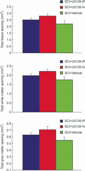
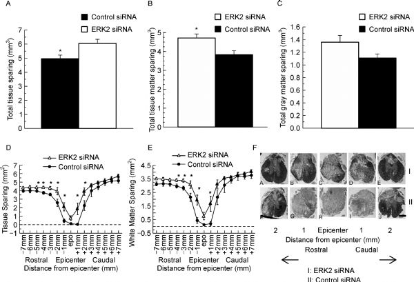
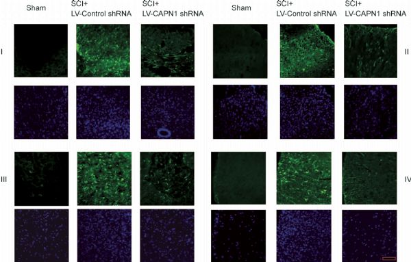
Abstract Neuronal damage, glial inflammation, and astrogliosis/astroglial scar formation are major secondary injury mechanisms that are significant contributors to functional deficits after spinal cord injury (SCI). The objectives of the study were to evaluate the distinct roles of ERK2 vs. ERK1/2 and ERK1/2-calpain 1−NF-κB signal transduction in the tissue damage and astrogliosis/astroglial scar formation following SCI in rats. RNAi approaches, pharmacological intervention (U0126), Western blot analysis, immunofluorescence analysis, and histological assessment were used to target ERK1/2-calpain 1-NF-κB signal transduction pathway for neuroprotection. Histological staining analysis demonstrated that selectively reducing pERK2 using ERK2 siRNA, but not inhibition of pERK1/2 with U0126, significantly reduced lesion volume and improved total tissue sparing, white matter sparing, and gray matter sparing in spinal cord two weeks after contusive SCI. An ERK1/2-calpain 1-NF-κB signal transduction pathway was involved in the astroglial scar formation after SCI. Blockade of ERK1/2 by U0126 decreased calpain 1 expression 4 h following SCI. Selective calpain 1 reduction by lentiviral shRNA attenuated astroglial NF-κB activity and astroglial scar formation after SCI in rats. Taken together, these results demonstrate the involvement of individual ERK2 and calpain 1 signaling pathways in tissue damage and astrogliosis/astroglial scar formation in animal models of SCI. Therefore, targeting individual ERK and its downstream signal transduction of calpain 1-NF-κB may provide greater potential as novel therapeutics for minimizing tissue damage and astroglial scar formation following SCI.
|
| Keywords
calpain 1
ERK1/2
RNAi
neurodegeneration
astrogliosis
spinal cord injury
|
|
Corresponding Author(s):
Chen Guang Yu
|
|
Just Accepted Date: 16 September 2015
Online First Date: 13 October 2015
Issue Date: 30 October 2015
|
|
| 1 |
Agrawal A, Dillon S, Denning T L, Pulendran B (2006). ERK1−/− mice exhibit Th1 cell polarization and increased susceptibility to experimental autoimmune encephalomyelitis. J Immunol, 176(10): 5788–5796
https://doi.org/10.4049/jimmunol.176.10.5788
pmid: 16670284
|
| 2 |
Aigner A (2006). Gene silencing through RNA interference (RNAi) in vivo: strategies based on the direct application of siRNAs. J Biotechnol, 124(1): 12–25
https://doi.org/10.1016/j.jbiotec.2005.12.003
pmid: 16413079
|
| 3 |
Borgens R B, Liu-Snyder P (2012). Understanding secondary injury. Q Rev Biol, 87(2): 89–127
https://doi.org/10.1086/665457
pmid: 22696939
|
| 4 |
Brambilla R, Hurtado A, Persaud T, Esham K, Pearse D D, Oudega M, Bethea J R (2009). Transgenic inhibition of astroglial NF-kappa B leads to increased axonal sparing and sprouting following spinal cord injury. J Neurochem, 110(2): 765–778
https://doi.org/10.1111/j.1471-4159.2009.06190.x
pmid: 19522780
|
| 5 |
Bramlett H M, Dietrich W D (2007). Progressive damage after brain and spinal cord injury: pathomechanisms and treatment strategies. Prog Brain Res, 161: 125–141
https://doi.org/10.1016/S0079-6123(06)61009-1
pmid: 17618974
|
| 6 |
Cargnello M, Roux P P (2011). Activation and function of the MAPKs and their substrates, the MAPK-activated protein kinases. Microbiol Mol Biol Rev, 75(1): 50–83
https://doi.org/10.1128/MMBR.00031-10
pmid: 21372320
|
| 7 |
Colak A, Kaya M, Karaoğlan A, Sağmanligil A, Akdemir O, Sahan E, Celik O (2009). Calpain inhibitor AK 295 inhibits calpain-induced apoptosis and improves neurologic function after traumatic spinal cord injury in rats. Neurocirugia (Astur), 20(3): 245–254
https://doi.org/10.1016/S1130-1473(09)70163-0
pmid: 19575128
|
| 8 |
Doshi S, Lynch D R (2009). Calpain and the glutamatergic synapse. Front Biosci (Schol Ed), 1(1): 466–476
https://doi.org/10.2741/s38
pmid: 19482714
|
| 9 |
Drake C R, Aissaoui A, Argyros O, Serginson J M, Monnery B D, Thanou M, Steinke J H, Miller A D (2010). Bioresponsive small molecule polyamines as noncytotoxic alternative to polyethylenimine. Mol Pharm, 7(6): 2040–2055
https://doi.org/10.1021/mp9002249
pmid: 20929266
|
| 10 |
Geddes J W, Saatman K E (2010). Targeting individual calpain isoforms for neuroprotection. Exp Neurol, 226(1): 6–7
https://doi.org/10.1016/j.expneurol.2010.07.025
pmid: 20682310
|
| 11 |
Hetman M, Gozdz A (2004). Role of extracellular signal regulated kinases 1 and 2 in neuronal survival. Eur J Biochem, 271(11): 2050–2055
https://doi.org/10.1111/j.1432-1033.2004.04133.x
pmid: 15153093
|
| 12 |
Ishikawa T, Suzuki H, Ishikawa K, Yasuda S, Matsui T, Yamamoto M, Kakeda T, Yamamoto S, Owada Y, Yaksh T L (2014). Spinal cord ischemia/injury. Curr Pharm Des, 20(36): 5738–5743
https://doi.org/10.2174/1381612820666140204113252
pmid: 24502574
|
| 13 |
Karimi-Abdolrezaee S, Billakanti R (2012). Reactive astrogliosis after spinal cord injury-beneficial and detrimental effects. Mol Neurobiol, 46(2): 251–264
https://doi.org/10.1007/s12035-012-8287-4
pmid: 22684804
|
| 14 |
Kim J Y, Park K J, Kim G H, Jeong E A, Lee D Y, Lee S S, Kim D J, Roh G S, Song J, Ki S H, Kim W H (2013). In vivo activating transcription factor 3 silencing ameliorates the AMPK compensatory effects for ER stress-mediated β-cell dysfunction during the progression of type-2 diabetes. Cell Signal, 25(12): 2348–2361
https://doi.org/10.1016/j.cellsig.2013.07.028
pmid: 23916985
|
| 15 |
Kuchay S M, Chishti A H (2007). Calpain-mediated regulation of platelet signaling pathways. Curr Opin Hematol, 14(3): 249–254
https://doi.org/10.1097/MOH.0b013e3280ef68f8
pmid: 17414215
|
| 16 |
Kwon B K, Sekhon L H, Fehlings M G (2010). Emerging repair, regeneration, and translational research advances for spinal cord injury. Spine, 35(21 Suppl): S263–S270
https://doi.org/10.1097/BRS.0b013e3181f3286d
pmid: 20881470
|
| 17 |
Li L, Wu Y, Wang C, Zhang W (2012). Inhibition of PAX2 gene expression by siRNA (polyethylenimine) in experimental model of obstructive nephropathy. Ren Fail, 34(10): 1288–1296
https://doi.org/10.3109/0886022X.2012.723662
pmid: 23078635
|
| 18 |
Liu J, Liu M C, Wang K K (2008). Calpain in the CNS: from synaptic function to neurotoxicity. Sci Signal, 1(14): re1
https://doi.org/10.1126/stke.114re1
pmid: 18398107
|
| 19 |
Liu L, Zhang R, Liu K, Zhou H, Tang Y, Su J, Yu X, Yang X, Tang M, Dong Q (2009). Tissue kallikrein alleviates glutamate-induced neurotoxicity by activating ERK1. J Neurosci Res, 87(16): 3576–3590
https://doi.org/10.1002/jnr.22151
pmid: 19598250
|
| 20 |
Lu P Y, Woodle M C (2008). Delivering small interfering RNA for novel therapeutics. Methods Mol Biol, 437: 93–107
https://doi.org/10.1007/978-1-59745-210-6_3
pmid: 18369963
|
| 21 |
Luo M C, Zhang D Q, Ma S W, Huang Y Y, Shuster S J, Porreca F, Lai J (2005). An efficient intrathecal delivery of small interfering RNA to the spinal cord and peripheral neurons. Mol Pain, 1(1): 29
https://doi.org/10.1186/1744-8069-1-29
pmid: 16191203
|
| 22 |
Nakazawa T, Shimura M, Ryu M, Nishida K, Pagès G, Pouysségur J, Endo S (2008). ERK1 plays a critical protective role against N-methyl-D-aspartate-induced retinal injury. J Neurosci Res, 86(1): 136–144
https://doi.org/10.1002/jnr.21472
pmid: 17722069
|
| 23 |
Rabchevsky A G, Fugaccia I, Sullivan P G, Blades D A, Scheff S W (2002). Efficacy of methylprednisolone therapy for the injured rat spinal cord. J Neurosci Res, 68(1): 7–18
https://doi.org/10.1002/jnr.10187
pmid: 11933044
|
| 24 |
Ray S K, Hogan E L, Banik N L (2003). Calpain in the pathophysiology of spinal cord injury: neuroprotection with calpain inhibitors. Brain Res Brain Res Rev, 42(2): 169–185
https://doi.org/10.1016/S0165-0173(03)00152-8
pmid: 12738057
|
| 25 |
Saatman K E, Creed J, Raghupathi R (2010). Calpain as a therapeutic target in traumatic brain injury. Neurotherapeutics, 7(1): 31–42
https://doi.org/10.1016/j.nurt.2009.11.002
pmid: 20129495
|
| 26 |
Schumacher P A, Siman R G, Fehlings M G (2000). Pretreatment with calpain inhibitor CEP-4143 inhibits calpain I activation and cytoskeletal degradation, improves neurological function, and enhances axonal survival after traumatic spinal cord injury. J Neurochem, 74(4): 1646–1655
https://doi.org/10.1046/j.1471-4159.2000.0741646.x
pmid: 10737623
|
| 27 |
Springer J E, Azbill R D, Kennedy S E, George J, Geddes J W (1997). Rapid calpain I activation and cytoskeletal protein degradation following traumatic spinal cord injury: attenuation with riluzole pretreatment. J Neurochem, 69(4): 1592–1600
https://doi.org/10.1046/j.1471-4159.1997.69041592.x
pmid: 9326288
|
| 28 |
Sribnick E A, Samantaray S, Das A, Smith J, Matzelle D D, Ray S K, Banik N L (2010). Postinjury estrogen treatment of chronic spinal cord injury improves locomotor function in rats. J Neurosci Res, 88(8): 1738–1750
pmid: 20091771
|
| 29 |
Tian D S, Yu Z Y, Xie M J, Bu B T, Witte O W, Wang W (2006). Suppression of astroglial scar formation and enhanced axonal regeneration associated with functional recovery in a spinal cord injury rat model by the cell cycle inhibitor olomoucine. J Neurosci Res, 84(5): 1053–1063
https://doi.org/10.1002/jnr.20999
pmid: 16862564
|
| 30 |
Wall E A, Zavzavadjian J R, Chang M S, Randhawa B, Zhu X, Hsueh R C, Liu J, Driver A, Bao X R, Sternweis P C, Simon M I, Fraser I D (2009). Suppression of LPS-induced TNF-alpha production in macrophages by cAMP is mediated by PKA-AKAP95-p105. Sci Signal, 2(75): ra28
https://doi.org/10.1126/scisignal.2000202
pmid: 19531803
|
| 31 |
Wittrup A, Lieberman J (2015). Knocking down disease: a progress report on siRNA therapeutics. Nat Rev Genet, 16(9): 543–552
https://doi.org/10.1038/nrg3978
pmid: 26281785
|
| 32 |
Wu J, Pajoohesh-Ganji A, Stoica B A, Dinizo M, Guanciale K, Faden A I (2012). Delayed expression of cell cycle proteins contributes to astroglial scar formation and chronic inflammation after rat spinal cord contusion. J Neuroinflammation, 9(1): 169
https://doi.org/10.1186/1742-2094-9-169
pmid: 22784881
|
| 33 |
Yiu G, He Z (2006). Glial inhibition of CNS axon regeneration. Nat Rev Neurosci, 7(8): 617–627
https://doi.org/10.1038/nrn1956
pmid: 16858390
|
| 34 |
Yu C G (2012). Distinct roles for ERK1 and ERK2 in pathophysiology of CNS. Front Biol, 7 (3): 267–276
|
| 35 |
Yu C G, Geddes J W (2007). Sustained calpain inhibition improves locomotor function and tissue sparing following contusive spinal cord injury. Neurochem Res, 32(12): 2046–2053
https://doi.org/10.1007/s11064-007-9347-4
pmid: 17476592
|
| 36 |
Yu C G, Li Y, Raza K, Yu X X, Ghoshal S, Geddes J W (2013). Calpain 1 knockdown improves tissue sparing and functional outcomes after spinal cord injury in rats. J Neurotrauma, 30(6): 427–433
https://doi.org/10.1089/neu.2012.2561
pmid: 23102374
|
| 37 |
Yu C G, Singh R, Crowdus C, Raza K, Kincer J, Geddes J W (2014). Fenbendazole improves pathological and functional recovery following traumatic spinal cord injury. Neuroscience, 256: 163–169
https://doi.org/10.1016/j.neuroscience.2013.10.039
pmid: 24183965
|
| 38 |
Yu C G, Yezierski R P (2005). Activation of the ERK1/2 signaling cascade by excitotoxic spinal cord injury. Brain Res Mol Brain Res, 138(2): 244–255
https://doi.org/10.1016/j.molbrainres.2005.04.013
pmid: 15922485
|
| 39 |
Yu C G, Yezierski R P, Joshi A, Raza K, Li Y, Geddes J W (2010). Involvement of ERK2 in traumatic spinal cord injury. J Neurochem, 113(1): 131–142
https://doi.org/10.1111/j.1471-4159.2010.06579.x
pmid: 20067580
|
| 40 |
Yuan Y M, He C (2013). The glial scar in spinal cord injury and repair. Neurosci Bull, 29(4): 421–435
https://doi.org/10.1007/s12264-013-1358-3
pmid: 23861090
|
| 41 |
Zhang S X, Underwood M, Landfield A, Huang F F, Gison S, Geddes J W (2000). Cytoskeletal disruption following contusion injury to the rat spinal cord. J Neuropathol Exp Neurol, 59(4): 287–296
pmid: 10759184
|
| 42 |
Zhao P, Waxman S G, Hains B C (2007). Extracellular signal-regulated kinase-regulated microglia-neuron signaling by prostaglandin E2 contributes to pain after spinal cord injury. J Neurosci, 27(9): 2357–2368
https://doi.org/10.1523/JNEUROSCI.0138-07.2007
pmid: 17329433
|
| 43 |
Zhuang S, Schnellmann R G (2006). A death-promoting role for extracellular signal-regulated kinase. J Pharmacol Exp Ther, 319(3): 991–997
https://doi.org/10.1124/jpet.106.107367
pmid: 16801453
|
|
Viewed |
|
|
|
Full text
|
|
|
|
|
Abstract
|
|
|
|
|
Cited |
|
|
|
|
| |
Shared |
|
|
|
|
| |
Discussed |
|
|
|
|